Revolutionizing Cancer Cell Analysis
A Custom Software Solution for High-Throughput Microscopy Imaging
| Industry: | Healthcare |
|---|---|
| Location: | Thailand |
| Platform: | Desktop application |

A Custom Software Solution for High-Throughput Microscopy Imaging
| Industry: | Healthcare |
|---|---|
| Location: | Thailand |
| Platform: | Desktop application |
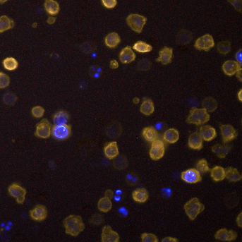
Our client, a pioneering startup composed of a team of scientists from Thailand and Germany, is at the forefront of cancer research, specializing in the detection of rare cancer cells via liquid biopsy method using advanced microscopy imaging. Despite their innovative approach, they faced significant operational hurdles with their existing tools.
Until recently, the team relied on the "Columbus Image Data Storage and Analysis System" , a robust platform developed by Revvity Inc, a publicly-traded company (NYSE: RVTY). However, the rigidity of Columbus, particularly its lack of customization options, significantly hampered their research. The system required approximately four hours of intensive labor by three scientists to process a single subject ID, a bottleneck that slowed critical advancements in their studies.
In their quest for a tailored solution, the startup had previously invested $30,000 in a freelance software developer. Unfortunately, this collaboration ended unfavorably without the delivery of any tangible software or source codes, leaving only a prototype demonstration video. This setback not only cost them financially but also left the team disheartened and cautious about future engagements.
Despite these challenges, the startup remained committed to improving their operational efficiency through technology. They envisioned a custom software solution that could significantly reduce their data processing time from four hours to less than one hour. Moreover, they believed that integrating artificial intelligence could further accelerate the process, potentially reducing analysis time to mere minutes. This efficiency gain was seen as critical to scaling their operations and enhancing their research capabilities.
Determined to turn their vision into reality, the startup sought a reliable software development partner who could understand their unique needs and deliver a high-quality, budget-friendly solution. This led them to our team, known for our expertise in custom software development and our commitment to transforming challenges into innovative technological solutions.






Our initial step involved a thorough investigation into the previous project's shortcomings. By engaging with contacts familiar with both the startup team and the previous freelancer, we pinpointed communication breakdown as the critical issue. It became evident that the freelancer had overpromised capabilities without delivering, and the startup team, despite their expertise in science, lacked clarity in defining software specifications. This misalignment had led to project failure.
Committed to avoiding past pitfalls, we initiated a series of deep-dive sessions to fully comprehend the startup's needs. This began with onsite visits to their laboratory in Thailand and was complemented by numerous online discussions. These sessions were crucial in transitioning from a high-level concept to a detailed and actionable project scope.
The project specifications, initially drafted by a German scientist, were steeped in specialized scientific terminology, making them challenging to translate into actionable software requirements. Recognizing this, we undertook a careful analysis to ensure nothing was lost in translation. This process involved multiple revisions of the user stories—approximately five iterations—until there was mutual clarity and agreement on the project scope.
Our collaborative approach resulted in a refined set of user stories that accurately reflected 95% of the startup's requirements. This document served as a foundational blueprint for the development process, ensuring all parties had a clear and shared understanding of the deliverables.
Understanding the dynamic nature of software development, especially in a field as complex as computational pathology, we established a Memorandum of Understanding (MOU). This MOU ensured both parties agreed to maintain a flexible, respectful, and transparent relationship. It outlined how changes could be managed, ensuring they were within reasonable efforts and aligned with the agreed-upon user stories and project scope.
After finalizing the user stories, our next pivotal discussion with the startup revolved around selecting the appropriate software format to meet their operational needs. Given their current reliance on the Columbus system—a web-based application operating within a local area network—we explored various options to optimize functionality while addressing their specific concerns.
| Internet Independence: |
The startup emphasized the necessity for a system that remains operational without
internet connectivity. Citing concerns over potential network failures, they required
a robust solution that ensures uninterrupted access to critical functionalities.
|
|---|---|
| Cost Efficiency: |
As a startup with limited financial resources, minimizing ongoing software licensing fees
was crucial. They sought a cost-effective solution that aligns with their budget constraints.
|
| Security Considerations: |
The security of their data and operations was a paramount concern, driving the need for a secure
platform that could safeguard sensitive research data.
|
| Technology Selection: |
Based on these criteria, we chose to develop a desktop application targeted for Windows platforms,
leveraging Python for its versatility and robustness. For the user interface, we selected PySide,
an open-source interface builder under LGPL, which offers the necessary flexibility without
the burden of licensing fees. This decision supported the startup's requirement for a secure,
reliable, and cost-effective software solution.
|
A significant enhancement in our solution was the integration of a proprietary AI library designed for high-precision cell identification. This enables the automated detection of cell events within images with exceptional accuracy. This technology not only streamlines the analysis process but also significantly reduces the time required for data processing, aligning perfectly with the startup’s goal to enhance efficiency in their research operations.
The culmination of these decisions and integrations resulted in a powerful, standalone desktop application that met all the startup's operational requirements and financial constraints, while also setting a new standard in accuracy and efficiency for their research processes.


